
 EN
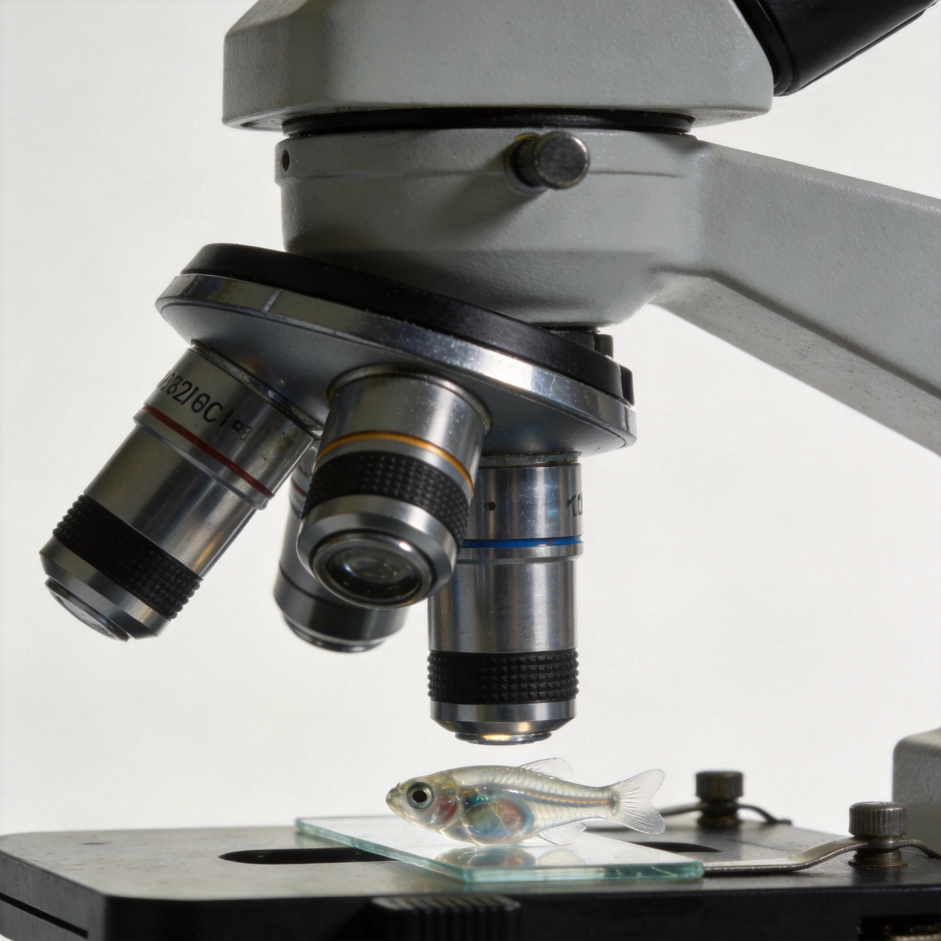
A facility upgrade is rarely about replacing a camera because it is old. It is about deciding whether the images your team collects are detailed, consistent, and usable enough for the questions now coming through the lab. A Zebrafish Imaging System can become a bottleneck when researchers need to distinguish subtle morphology, follow fluorescence over time, compare cohorts, or return to an image months later and understand how it was acquired.
For a core facility, university lab, or screening group, a high-resolution upgrade should be assessed as a workflow decision. The relevant question is not simply “how many pixels?” It is whether the system gives users dependable visual evidence, repeatable acquisition settings, practical throughput, and an analysis path that fits the facility’s staffing and data-management capacity.
When lower image detail starts to limit the research question
Zebrafish larvae are optically accessible during early development, which is one reason live imaging can be so informative. But that advantage only helps when the optical setup, sample preparation, and acquisition settings are matched to the feature being studied. A borderline structure may look like biological variation in one image and background in another. If different operators compensate with different exposure, focus, or magnification choices, the facility may create avoidable ambiguity before analysis even begins.
That is usually the first signal that an upgrade deserves discussion: the team spends more time debating whether a feature is real than extracting a measurement from it. A high-resolution Zebrafish Imaging System can help make fine anatomical boundaries, fluorescent signal distribution, and phenotype documentation easier to assess, provided its resolution is appropriate for the biological endpoint. Higher resolution alone is not automatically useful if it brings longer capture times, more light exposure, or files the facility cannot manage.
What an upgrade can change for a shared facility
Shared imaging environments have a different challenge from a single-user bench. A system must support repeatable work across people, projects, and time. When users can save acquisition recipes, document objective and illumination choices, and retrieve image metadata with the dataset, a facility is in a stronger position to compare runs rather than relying on memory or handwritten notes. That matters because standardized reporting and metadata are increasingly important themes in zebrafish research reproducibility.
Resolution also affects how confidently a facility can support multiple application types. A group studying development may need clear morphology; another may need fluorescence-based spatial patterns; a third may be screening for a change that is modest but biologically meaningful. The practical advancement is not a claim that one platform solves every assay. It is the ability to define what each assay requires and configure a repeatable imaging workflow around it.
Evaluate the full imaging workflow, not the specification sheet alone
Before approving an upgrade, map the journey from live fish to a usable result. The imaging head is only one part of that journey. Sample positioning, focus stability, illumination control, plate or chamber compatibility, software export, storage, and analyst review all affect the final dataset.
- Start with the endpoint. List the smallest feature, intensity difference, or motion event that users need to resolve. This establishes a meaningful resolution requirement.
- Check acquisition consistency. Ask whether users can retain and reuse settings for objective, exposure, illumination, frame rate, and sample orientation.
- Assess live-sample handling. A workflow that requires excessive repositioning or prolonged light exposure may reduce the practical value of image detail.
- Plan for analysis. Confirm that exported files, metadata, and naming conventions can move into the facility’s existing review or quantification process.
- Test representative assays. A vendor demonstration should include the strains, developmental stages, labels, and endpoints that matter to your users—not only visually striking examples.
This approach helps a facility avoid buying more optical capability than it can deploy. It also reveals where a modest configuration change, staff training, or an SOP update may be as important as the instrument itself.
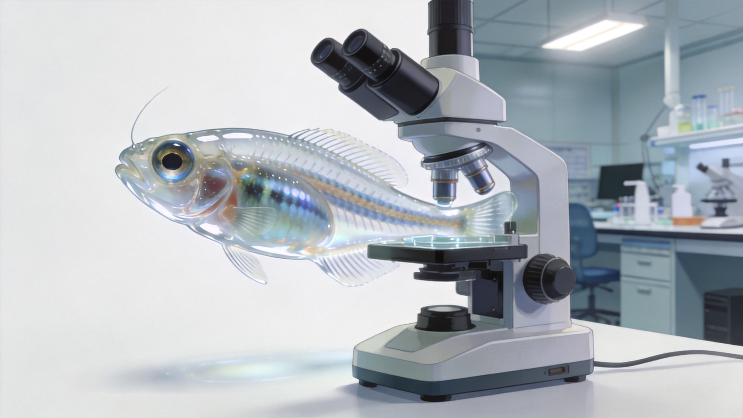
High-resolution imaging supports decisions when it is paired with standardization
A frequent pain point in multi-user labs is operator-dependent scoring. One researcher may accept a slightly soft image; another may repeat the capture; a third may apply a different threshold during review. Over a multi-week study, those small choices can make pooled data harder to interpret. The published protocol might look complete, but in a real facility, users often need a local version that accounts for the available optics, room conditions, mounting method, and approved analysis workflow.
A high-resolution Zebrafish Imaging System can support a better local standard when the upgrade includes practical controls: preset acquisition methods, a short qualification image set, routine focus and illumination checks, and a simple metadata checklist. For example, a facility might record strain, developmental stage, objective, exposure, filter set, mounting method, operator, and acquisition date alongside the file. These are not bureaucratic extras; they make later comparison more credible.
Balance resolution with throughput, light management, and storage
More detail increases data volume, and more frames can extend acquisition time. That trade-off needs a deliberate decision. For a high-content screen, throughput and consistent automated capture may matter more than extracting the maximum possible detail from every larva. For a developmental or validation study, the facility may choose slower acquisition at selected time points because the added detail answers a defined question.
Light management deserves the same attention. Live imaging can be gentle when illumination and exposure are chosen thoughtfully, yet unnecessary intensity or repeated imaging can complicate a sensitive experiment. During an evaluation, ask the team to compare image quality at the lowest practical illumination and exposure settings for the assay. The right Zebrafish Imaging System is the one that helps the facility obtain interpretable images under conditions its users can reproduce.
Build the business case around usable capacity
Procurement teams often need more than a scientific rationale. A durable proposal explains who will use the system, which current delays it addresses, what training is required, and how service or application support will fit the facility. Hunter Lab focuses its product line on zebrafish instruments and applications, which can be relevant when a facility values zebrafish-specific technical discussion. Still, the selection should be based on documented fit for the facility’s own assays, budget process, and support expectations.
Use a short acceptance plan before purchase: define representative samples, success criteria for image detail, expected capture time, expected file workflow, and the people who will approve results. Then compare the proposed configuration against the current process. This gives scientists, facility managers, and purchasing stakeholders a common evidence base instead of a conversation driven only by headline specifications.
A practical upgrade checklist
- Define the biological features and measurements that current images do not support reliably.
- Document present bottlenecks in capture, review, repeat imaging, and data handoff.
- Specify acceptable acquisition consistency and metadata requirements for shared use.
- Evaluate image detail using your own representative samples and endpoints.
- Check whether throughput, live-sample handling, and storage remain workable at the selected settings.
- Plan SOPs, operator training, qualification checks, and a service path before the system enters routine use.
Make the upgrade a research-capability decision
Upgrading to a high-resolution Zebrafish Imaging System is justified when it removes a defined barrier between the specimen and a defensible result. The value comes from combining appropriate optical detail with stable acquisition, manageable workflow, and records that let future users understand what happened in each run. For facilities that serve several research groups, that combination can turn imaging from a variable handoff into a shared, repeatable research capability.
Begin with the assays your users already struggle to document, set acceptance criteria around those assays, and evaluate the workflow end to end. That is a more reliable path to an imaging advancement than choosing a system on resolution alone.








