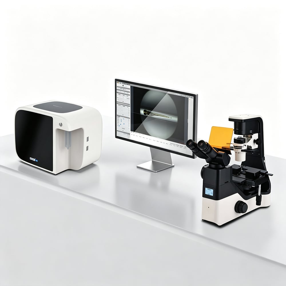
What Is a Zebrafish Panoramic Imaging System?
A laboratory imaging system designed for zebrafish research captures, processes, and analyzes images of live zebrafish larvae across early developmental stages. Unlike generic life science imagers, a zebrafish-specific imaging system is engineered to work with the unique optical and physiological properties of larvae at 3–7 days post-fertilization (dpf)—the window when cardiovascular, morphological, and behavioral phenotypes are most informative and accessible.
The key biological advantage that makes optical imaging systems so effective for zebrafish research is larval transparency. Zebrafish are optically transparent during early development, making them uniquely suited for non-invasive in vivo imaging without surgical preparation, anesthesia for tissue access, or radioisotope labeling. This property means that a well-configured imaging system can deliver high-quality structural and functional data directly through the intact animal—no ex vivo preparation required. For labs evaluating a zebrafish imaging platform for the first time, this transparency-driven optical access is the foundational reason the model system works so efficiently with optical hardware.
Key Product Characteristics
The Zebrafish Panoramic Imaging System in this category is built for 3–7 dpf larval imaging with a focus on throughput consistency, image quality, and multi-modal coverage across both brightfield and fluorescence channels:
-
Automated sample loading: Reduces inter-operator variability by standardizing larval positioning across imaging runs. In practice, inconsistent loading is one of the most common sources of avoidable noise before a single image is captured—automated handling addresses this at the workflow level rather than relying on operator technique.
-
Multi-angle brightfield imaging: Captures panoramic views of larval morphology from multiple orientations in a single automated session, eliminating the need for manual repositioning and enabling complete external morphology documentation.
-
Fluorescence imaging compatibility: Compatible with standard transparent and optically-accessible zebrafish strains (e.g., casper, crystal) for clear cardiac and vascular visualization without pigment interference. This enables GFP-reporter cardiac studies and fluorescence-coupled phenotyping—including vascular imaging relevant to labs also exploring zebrafish blood flow analysis—alongside standard brightfield runs.
-
High-resolution brightfield capture: High-resolution brightfield imaging captures fine cardiac structural details including atrial-ventricular junction and pericardial space morphology (the space between the heart and body wall, which expands visibly in pericardial edema phenotypes) alongside functional measurements.
-
Full 3–7 dpf developmental stage coverage: Designed for the early larval window, covering the complete range of cardiovascular and morphological development from initial cardiac looping through chamber maturation.
Common Applications
Zebrafish imaging systems serve a broad range of research, safety testing, and screening applications across academic, pharmaceutical, and industrial customer segments:
-
Cardiovascular phenotyping: Morphological assessment of cardiac structure—chamber size, pericardial edema, and vascular patterning—is one of the most common uses of panoramic brightfield imaging. When you look at a brightfield image of a 5 dpf larval heart, you can directly observe pericardial space changes and chamber morphology—not just infer them from a numerical summary.
-
Developmental toxicology and teratogenicity screening: Pharmaceutical R&D teams and cosmetics safety labs use zebrafish imaging to evaluate the morphological effects of compound exposure across the 3–7 dpf window. Combined morphological and functional phenotyping in the same session reduces animal use and experimental time per compound.
-
Blood flow and vascular analysis: When combined with fluorescent reporter lines, fluorescence-compatible imaging systems can visualize circulatory dynamics non-invasively—relevant for labs running vascular phenotyping alongside cardiac assessments.
-
Multi-stage developmental studies: The same developmental parameter behaves differently at 3 dpf compared to 5 dpf—and if your assay does not hold developmental stage constant across groups, you risk comparing larvae that are not physiologically equivalent. Imaging systems covering the full 3–7 dpf range with consistent acquisition settings support more reliable longitudinal and cross-stage comparisons.
-
Safety testing for cosmetics, nutraceuticals, and pharmaceutical screening: Labs in these segments value imaging systems that generate reproducible, publication-ready output. A pharmaceutical R&D team attempting to replicate a published zebrafish cardiotoxicity protocol using inadequate imaging equipment frequently finds that image quality is insufficient to extract the reported quantitative endpoints—an avoidable procurement failure when the right imaging specifications are confirmed before purchase.
How to Choose the Right Imaging System
When evaluating an imaging system for institutional or grant-funded procurement, the decision should hinge on a realistic assessment of your research program's actual requirements. Here is a structured framework for the most important decision points:
-
What imaging modalities do you need now, and what might you need within two years? Zebrafish cardiovascular and morphological research programs often expand their modality requirements as they mature—initial brightfield characterization is typically followed by fluorescence for reporter lines or vascular imaging. Procuring a system that supports multi-modal imaging from the start avoids a forced platform transition within the first equipment lifecycle.
-
What is your actual throughput requirement? Automated loading and multi-angle capture reduce per-larva imaging time, but the throughput benefit only matters relative to your screen volume. For small exploratory studies, simpler setups may be adequate; for programs running regular batch imaging sessions, automation reduces both time cost and operator-driven variability.
-
How will you validate image quality for your specific assay? Request assay-specific performance data for the endpoints you plan to measure—not just general resolution or camera specifications. A vendor that can provide only generic specification sheets (rather than application-specific validation) makes it difficult to confirm whether the system will actually meet your experimental needs before purchase.
-
More parameters are not always better data: The instinct to maximize the number of imaging endpoints in a screen is understandable, but beyond a certain point, the analytical and statistical burden of multiple parameters starts to outweigh the added biological insight. Define the minimum endpoint set that answers your research question before selecting an imaging configuration.
-
Consider installation and post-sale support logistics: For labs evaluating vendors across geographies, installation responsiveness and local service coverage matter. Questions about zebrafish endurance testing device installation timelines and post-sale support response are worth raising with vendors before finalizing a purchase—especially for core facility procurement where instrument uptime is operationally critical.
Why Specialized Zebrafish Imaging Systems Matter for Research Labs
The zebrafish instrument market is served by a mix of large general life science equipment companies and manufacturers that specialize exclusively in zebrafish research platforms. The key distinction between these supplier types is depth of application focus: a vendor whose R&D resources are concentrated on zebrafish imaging specifically can provide deeper protocol guidance, assay-specific technical support, and more relevant product development than a diversified vendor managing hundreds of product lines simultaneously.
For international customers—including university labs in Asia, Europe, and North America—evaluating imaging systems from manufacturers with established export logistics experience, the relevant procurement questions include consistency of export documentation, international delivery coordination, and post-sale support capability for the destination region. Labs in these markets have established procurement channels for zebrafish research equipment, and experienced vendors provide the compliance documentation and delivery coordination that institutional purchase processes require.
Whether your lab is establishing a new zebrafish imaging capability, upgrading from an older platform, or coordinating a broader equipment plan that includes incubation infrastructure or other zebrafish research tools, the right imaging system selection depends on aligning the platform's capabilities with your actual assay requirements and support expectations—not on maximizing specification numbers alone.

 EN