
 EN
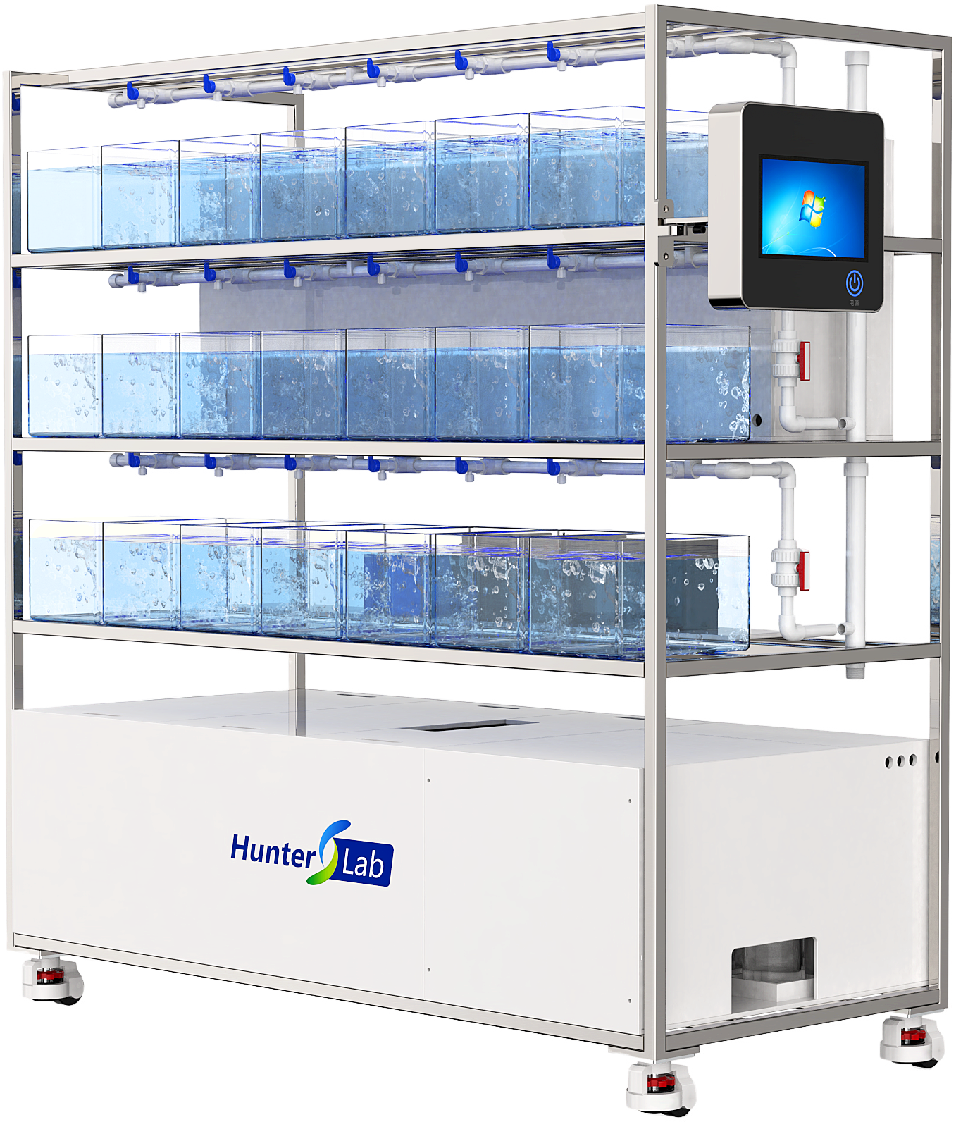
Why does a zebrafish imaging system with automated multi-well scanning save over 70% of your screening time?
In a screening lab, the slow part is rarely the camera exposure alone. It is the repeated sequence around it: placing a plate, finding each well, deciding whether the larva is positioned acceptably, setting focus, naming files, and returning later to score results. A Zebrafish Imaging System with automated multi-well scanning changes that sequence into a repeatable plate workflow. Under a well-defined comparison, that can remove more than 70% of hands-on screening time. It is not a universal promise; it is a workflow result that depends on the manual baseline, plate format, assay, and the settings used.
For teams moving from small pilot studies to high-throughput work, the practical question is not “Is automation faster?” It is “Which minutes disappear, which minutes remain, and do we still trust the data?” That distinction makes a purchasing claim testable instead of promotional.
Where manual screening loses time
A manual plate run contains many short decisions that add up. The operator moves from well to well, centers the subject, adjusts illumination or focus, starts acquisition, checks that the file was saved, and then repeats. Even when each action seems quick, a 96-well screen turns it into a long chain of context switches. A published automated heartbeat-analysis workflow notes that screening a 96-well plate can take roughly 30–50 minutes depending on the video length collected for each heart. That is useful as a scale reference, not as a specification for every assay.
The hidden cost is the variability in those choices. One operator may rescan borderline wells immediately; another may flag them for later. One may choose a slightly different focal plane. In a time-saving workflow, fewer repeated choices matter as much as fewer clicks, because a consistent capture sequence makes later review more comparable.
What automated multi-well scanning actually automates
Multi-well scanning means the platform follows a predefined map across the plate and applies the same acquisition recipe to the selected wells. Depending on the assay, that recipe can coordinate stage movement, autofocus or fixed-focus positions, illumination, image or video capture, file naming, and queueing for analysis. The important point is that automation converts a series of manual micro-decisions into parameters that can be reviewed before the run.
- Plate navigation: the system moves through wells in a defined order rather than relying on manual repositioning.
- Repeatable acquisition: exposure, frame count, and capture timing can be applied consistently across a batch.
- Structured data handling: well identity and acquisition settings travel with the file, reducing reconciliation work later.
- Exception-based review: staff spend their attention on wells that fail a quality rule or show a phenotype, not on every routine capture.
That last point is often where the largest gain appears. The goal is not to eliminate scientific judgment. It is to reserve judgment for exceptions, assay development, and biological interpretation.
How a 70% reduction can be realistic
Use a simple time budget. Suppose a manual 96-well run takes 90 minutes of active operator time: 45 minutes for positioning and capture, 20 minutes for file handling and review, and 25 minutes for interruptions, rescans, and recordkeeping. An automated Zebrafish Imaging System might reduce the active portion to a 10-minute setup, 10 minutes of exception review, and 5 minutes of run closeout while acquisition proceeds unattended. That is 25 minutes of active time rather than 90—about a 72% reduction.
The comparison is valid only when both paths collect equivalent data. If the automated workflow uses shorter videos, fewer fields, or a different quality threshold, the apparent gain is not an apples-to-apples result. Good teams therefore define the benchmark before a purchase evaluation: plate type, developmental stage, imaging mode, wells included, video length, rescan rules, and whether “screening time” means elapsed instrument time, hands-on time, or time to a review-ready dataset.
Why speed can improve consistency rather than compromise it
Automation is sometimes treated as a trade-off: more wells per day, less careful imaging. That is the wrong comparison. In a validated protocol, standardizing the acquisition path can reduce operator-to-operator variation. For brightfield or fluorescence phenotyping, the relevant control is not simply a faster stage; it is whether the system applies the same imaging conditions, preserves plate and well metadata, and makes outliers visible for review.
Zebrafish larvae are particularly suitable for optical workflows during early development, but that does not remove assay controls. Temperature, anesthesia, orientation, developmental stage, and motion all affect what the camera sees. If the imaging room varies substantially, timing and physiological readouts need to be interpreted against a documented baseline. Automation makes these conditions easier to record; it does not make them irrelevant.
Build the workflow around exceptions, not ideal wells
For a screening team, a useful acceptance rule has two parts. First, define what a successful well looks like: adequate focus, usable signal, correct plate position, and sufficient frames. Second, decide what happens when a well fails. It may be rescanned automatically, queued for a technician, or excluded according to a pre-approved rule. Without that exception path, a fast scanner can simply move the bottleneck into manual cleanup.
A practical validation run should compare manual and automated batches using the same plate layout. Track:
- hands-on minutes per plate;
- elapsed time to a complete image set;
- percentage of wells needing rescans;
- percentage of files correctly linked to plate and well identifiers; and
- agreement of the biological endpoint between workflows.
If throughput is the binding constraint, automated scanning reduces the bottleneck by addressing repeated positioning and routine capture—not by asking the operator to accept lower-quality data. That is also why efficiency should be measured as usable datasets per staff hour, not simply wells visited per hour.
Questions to ask when evaluating a zebrafish imaging system
A high-throughput demonstration should be specific enough for your own protocol. Ask the supplier or core-facility team to show the exact plate type and imaging mode you use. Confirm whether the system supports your planned field count and video duration, how focus behavior is handled, which metadata are retained, and how failed wells are flagged. Also ask which steps remain manual: loading, orientation checks, quality review, and image analysis may all have different ownership.
For cardiac or functional assays, do not stop at a scan-speed figure. Verify that the acquisition settings are compatible with the temporal resolution and signal quality needed for the endpoint. A rapid run that produces ambiguous videos is not time-saving; it creates a downstream annotation problem.
The takeaway
An automated Zebrafish Imaging System earns a “70%+ time-saving” claim when it removes most of the repeatable handling, navigation, capture, and recordkeeping from a baseline that previously required constant operator attention. The most useful evidence is your own controlled benchmark: same assay, same plate, same decision rules, and a clear separation between unattended acquisition time and staff time. When multi-well scanning is paired with exception-based review and robust metadata, the gain is more than speed—it is a screening workflow that is easier to scale, audit, and reproduce.








