
 EN
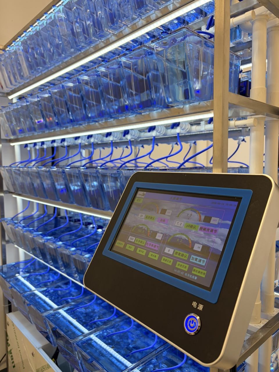
What imaging modalities are used in a zebrafish phenotype analysis system?
A Zebrafish Phenotype Analysis System is not defined by one camera or one assay. It is a workflow that connects sample handling, optics, image acquisition, and measurement to a biological question. In practice, the most useful modality is the one that makes the endpoint visible with enough consistency to compare control and treated groups. For a researcher planning a new assay, that usually means starting with the phenotype—not with the most sophisticated instrument.
Brightfield and fluorescence are the two workhorse channels in many Zebrafish Phenotype Analysis System workflows. Phase contrast, transmitted-light variants, confocal fluorescence, and specialized label-free methods may be added when the endpoint needs more contrast, depth information, or chemical specificity. The right combination depends on whether the study asks about morphology, movement, a reporter signal, tissue architecture, or a more specialized molecular feature.
Start with the phenotype you need to measure
Before selecting optics, write down the observable decision the assay must support. A Zebrafish Phenotype Analysis System for embryo morphology may need reliable whole-fish outlines, eye area, yolk-sac appearance, body length, spinal curvature, edema, or hatching status. A vascular or immune-cell assay may instead need a fluorescent reporter channel. Cardiac, behavioral, or developmental endpoints can require a short time series, repeated time points, or carefully controlled orientation.
This endpoint-first step prevents a common planning error: collecting visually impressive images that do not yield a stable measurement. Ask four practical questions: What signal will distinguish groups? Is it visible without a label? Does it occur near the surface or through tissue depth? How many fish and time points must the workflow process? The answers narrow the imaging choice quickly.
Brightfield imaging for whole-organism morphology
Brightfield imaging is often the baseline modality because it captures general anatomy without requiring fluorescent labeling. It is well suited to whole-embryo or larval observations such as gross developmental delay, body shape, pigmentation changes, eye size, tail curvature, pericardial edema, yolk-sac changes, and survival-related morphology. It is also practical for establishing orientation and locating the fish before other channels are acquired.
The appeal of brightfield is not that it answers every question. Its strength is rapid, interpretable imaging across many wells when the phenotype has visible contrast. Consistent illumination, focus strategy, plate choice, and larval orientation matter as much as raw camera resolution. If edges or internal structures are poorly separated, the workflow may benefit from color brightfield, transmitted-light adjustments, or a complementary channel rather than simply increasing magnification.
Fluorescence imaging for reporter-defined biology
Fluorescence imaging becomes valuable when the phenotype is tied to a labeled cell population, organ, pathway activity, or probe. Transgenic reporters and compatible stains can make vessels, immune cells, neurons, lipid deposits, oxidative-stress responses, or other selected structures easier to quantify than they would be in brightfield. A Zebrafish Phenotype Analysis System can use one fluorescence channel for a single target or multiple channels when the biological question requires comparison of distinct signals.
Widefield labeled-signal imaging is often a sensible screening choice when the goal is total signal, area, count, intensity distribution, or another measurement that does not demand fine depth separation. It can work alongside brightfield in the same acquisition plan: the transmitted-light channel supplies the whole-animal context, while fluorescence identifies the labeled biology. The study design should define exposure, gain, filter sets, background handling, and signal thresholds before comparing plates. Otherwise, differences in imaging conditions can be mistaken for phenotype differences.
Confocal imaging when depth and localization matter
Confocal fluorescence microscopy is useful when a widefield image combines signals from different depths and obscures the structure that matters. In this setting, optical sectioning can help researchers inspect localized features, reconstruct a region from a z-stack, or distinguish signals that overlap in a thicker sample. This makes confocal imaging a strong follow-up modality for selected wells or detailed mechanistic studies.
That added detail carries a throughput trade-off. More z-planes, channels, and fields of view increase acquisition time and data volume. Confocal imaging is therefore not automatically the first-line choice for every phenotype screen. A practical workflow often uses transmitted-light or widefield labeled-signal imaging to identify candidate phenotypes, then applies confocal imaging to a smaller, preplanned subset where depth-resolved evidence will change the interpretation.
Phase contrast, time-lapse, and specialized options
Phase contrast or related contrast-enhancing approaches can help where unlabeled structures have limited brightfield contrast. Time-lapse imaging is relevant when the phenotype is a process rather than a single appearance—for example, rhythmic movement, developmental progression, circulation-related motion, or behavioral change. In these cases, the acquisition plan needs a sampling schedule that captures the event without creating unnecessary light exposure or unmanageable file volumes.
Some studies use specialized label-free modalities, including Raman-based approaches, to investigate biochemical information. These methods can be valuable in dedicated research settings, but they are not a default requirement for routine phenotype analysis. The modality should earn its place by answering an endpoint that transmitted-light, reporter-channel, or confocal imaging cannot answer adequately within the required throughput.
How to choose the imaging modality
| Primary question | Useful starting modality | What to confirm during setup |
|---|---|---|
| Is overall development or morphology altered? | Brightfield | Focus, orientation, illumination, and whole-fish coverage |
| Is a labeled tissue, cell type, or reporter changing? | Widefield fluorescence | Channel separation, background, exposure consistency, and controls |
| Does the result depend on tissue depth or fine localization? | Confocal optical-section imaging | Z-step spacing, region definition, throughput, and data handling |
| Does the phenotype change over time? | Time-lapse transmitted-light or reporter-channel imaging | Frame interval, environmental stability, and motion handling |
For most teams, the most robust phenotype-analysis workflow is a layered one rather than a single-modality purchase decision. Use brightfield to establish morphology and context; add fluorescence when the biological signal is reporter-defined; reserve confocal imaging for depth-resolved questions. This sequence keeps the assay connected to an endpoint, a comparison group, and a repeatable measurement.
Build an acquisition plan before scaling
Run a small pilot before committing to a full plate. Include untreated or vehicle controls, a consistent developmental window, and a clear naming convention for wells, channels, and time points. Review a few images with the people who will analyze the data. Can they identify the phenotype using predefined criteria? Does the chosen workflow create a measurement that remains valid when different operators repeat the run?
The final choice is rarely “brightfield versus fluorescence” in the abstract. It is a match between biological signal, imaging modality, plate scale, and analysis method. By defining that match early, researchers can choose imaging that is informative, scalable, and easier to defend when phenotype results are compared across experiments.








