
 EN
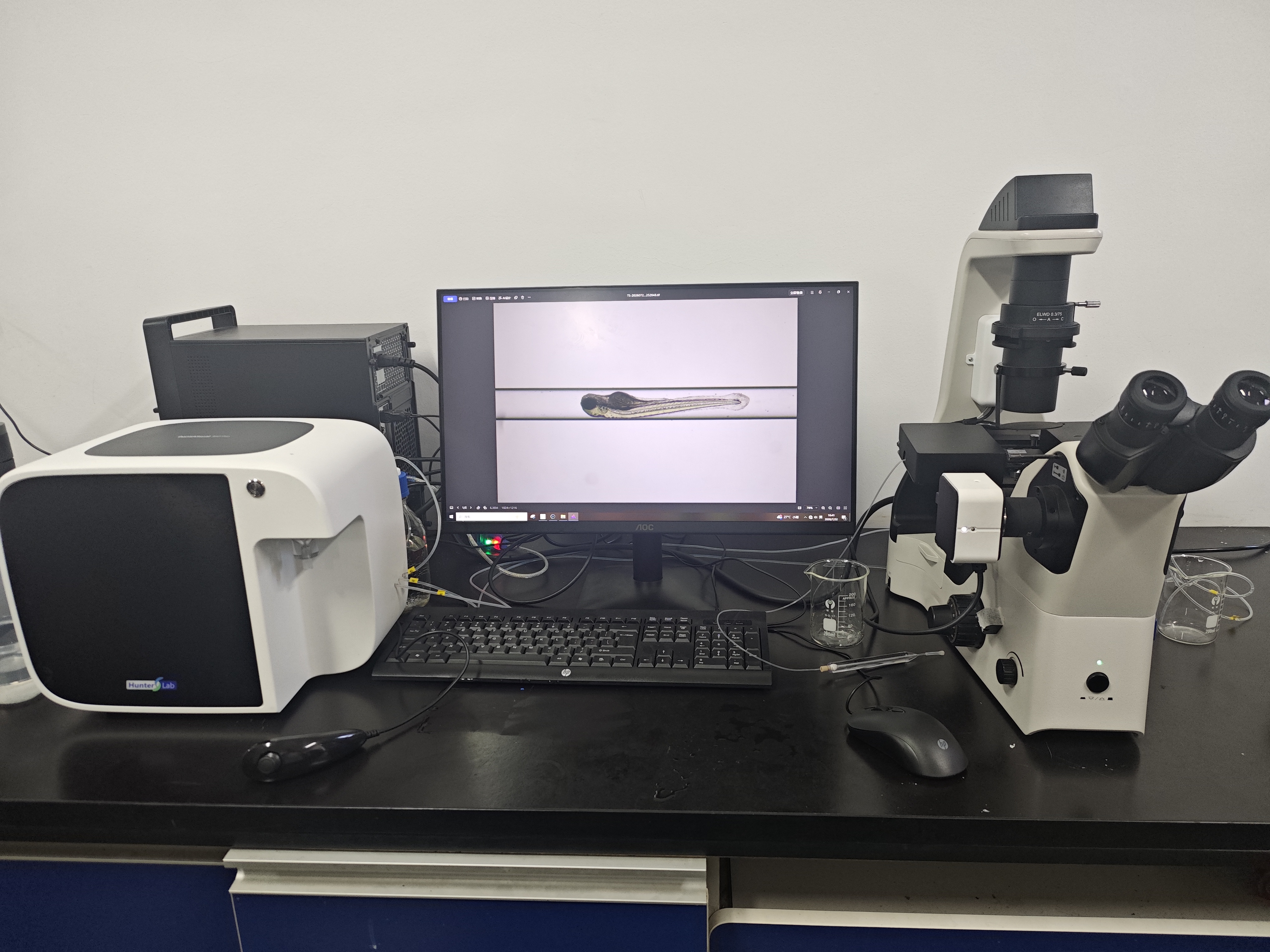
How does a zebrafish phenotype analysis system identify morphological abnormalities?
A Zebrafish Phenotype Analysis System does not replace biological judgment with a single image. It creates a controlled chain of evidence: embryos or larvae are prepared under a defined protocol, imaged consistently, measured against reference expectations, and reviewed before a morphology call is accepted. That sequence matters when a laboratory needs to distinguish a genuine developmental signal from ordinary variation in orientation, illumination, or developmental timing.
For academic groups, pharmaceutical teams, and cosmetics or health-product researchers, phenotype screening is valuable because it observes the organism as a whole. A structured workflow makes the resulting observations easier to compare across wells, plates, operators, and experimental days.
Start with a reference frame, not an isolated fish image
The first task of a Zebrafish Phenotype Analysis System is to make images comparable. Labs define the developmental stage, exposure window, housing conditions, anesthetic approach where applicable, imaging magnification, and plate layout before the run begins. Controls are acquired in the same session so that the analysis has a relevant baseline rather than an abstract “normal” image.
Image acquisition then aims for stable focus, illumination, and field of view. Depending on the assay, samples may be positioned so that a lateral or dorsal view exposes the anatomical features of interest. Some automated embryo-imaging workflows have been designed to work despite differences in embryo orientation, but consistency still improves downstream measurements. A useful screening record also keeps the plate, well, specimen identifier, time point, and exposure group connected to every image.
How imaging becomes measurable morphology
After capture, the Zebrafish Phenotype Analysis System separates the specimen from the background and locates usable anatomical landmarks. This can include the outer contour, head and trunk region, eye area, yolk sac, tail axis, pericardial region, or other assay-defined structures. Registration tools reported for zebrafish embryos can align an outer contour and internal regions so that measurements are taken in comparable locations rather than at arbitrary pixels.
The system converts the image into features that can be inspected or scored. Common morphology-oriented feature groups include:
- Body form: body length, curvature, tail bending, symmetry, and outline irregularity.
- Developmental landmarks: relative eye size, head shape, somite appearance, and timing-related changes visible at the selected stage.
- Localized changes: yolk-sac edema, pericardial edema, pigment distribution, tissue opacity, or altered organ-region shape when the assay supports that observation.
- Viability-related observations: coagulation, failed hatching at a defined observation point, or severe loss of normal posture.
Not every study needs every feature. The protocol should state which endpoints are relevant before screening begins. That prevents a broad imaging dataset from becoming an unfocused search for differences after the fact.
Detection means comparison against defined criteria
Detection is the comparison step. The Zebrafish Phenotype Analysis System can compare each specimen’s features with control distributions, reference annotations, threshold rules, or a trained classification model. A marked deviation may generate a flag for morphological abnormalities; it is not automatically a biological conclusion.
For example, a system may flag an embryo when its body axis departs from an acceptable curvature range, when the segmented yolk region is larger than the control reference, or when a landmark cannot be found with sufficient confidence. In model-based workflows, a classifier may return a normal/abnormal label or probabilities for pre-defined phenotype categories. Low-confidence outputs should be handled as review cases, not quietly forced into a category.
This distinction is important in high-throughput screening. Automated recognition can make the first pass faster and more consistent, while the criteria remain transparent enough for an investigator to understand why a sample was flagged.
Why human review remains part of the workflow
Historically, morphology scoring has relied heavily on visual assessment by an individual. A modern Zebrafish Phenotype Analysis System can reduce repetitive manual work, yet expert review remains essential for artifacts, borderline samples, and findings that would change an experimental decision. A bubble, shadow, partial occlusion, or poor focus can look like a phenotype to an algorithm if quality controls are weak.
Good practice is to queue flagged images with the original image, overlay or segmentation mask, measured features, confidence value, and the applicable scoring rule. Reviewers can then confirm, reject, or mark the specimen as ungradable. When possible, blinded review and a documented adjudication rule help prevent treatment knowledge from shaping the final call.
Build quality control into phenotype screening
Reliable imaging and detection depend on quality gates before and after analysis. Before measurement, a Zebrafish Phenotype Analysis System should identify images that are out of focus, overexposed, incomplete, or missing a specimen. During analysis, the laboratory should monitor control behavior, feature-distribution shifts, and the rate of ungradable images. After review, the team should retain enough metadata to trace every abnormality call back to its plate, well, image, and scoring version.
These checks also protect against a common problem in multicohort work: a small shift in camera settings or developmental timing can look like a treatment effect. Comparing contemporaneous controls and documenting protocol changes make the screening result more defensible.
A practical interpretation sequence
- Define phenotype endpoints and acceptance criteria before exposure or imaging.
- Acquire control and test images under the same documented conditions.
- Run image-quality checks and exclude ungradable images by rule.
- Extract landmarks and morphology features relevant to the assay.
- Use thresholds or validated models to flag candidate abnormalities.
- Review uncertain and high-impact calls with the image evidence and metadata.
- Summarize incidence, severity, and exclusion reasons at the group level.
What to look for when evaluating a system
When selecting a Zebrafish Phenotype Analysis System, researchers should ask whether the workflow supports their exact developmental stage and phenotype endpoints, preserves original images and metadata, exposes the scoring logic, and allows reviewers to inspect flagged cases. It is also worth asking how the system handles orientation, inconsistent image quality, and updates to a classification model. A well-designed system supports repeatable decisions; it should not hide uncertainty behind an automated label.
In short, morphological detection works as an evidence pipeline. Standardized imaging produces comparable records, feature extraction turns anatomy into measurable observations, and transparent review confirms whether a signal is meaningful. That combination gives phenotype screening a stronger foundation for follow-up biology, safety assessment, or experimental prioritization.








