
 EN
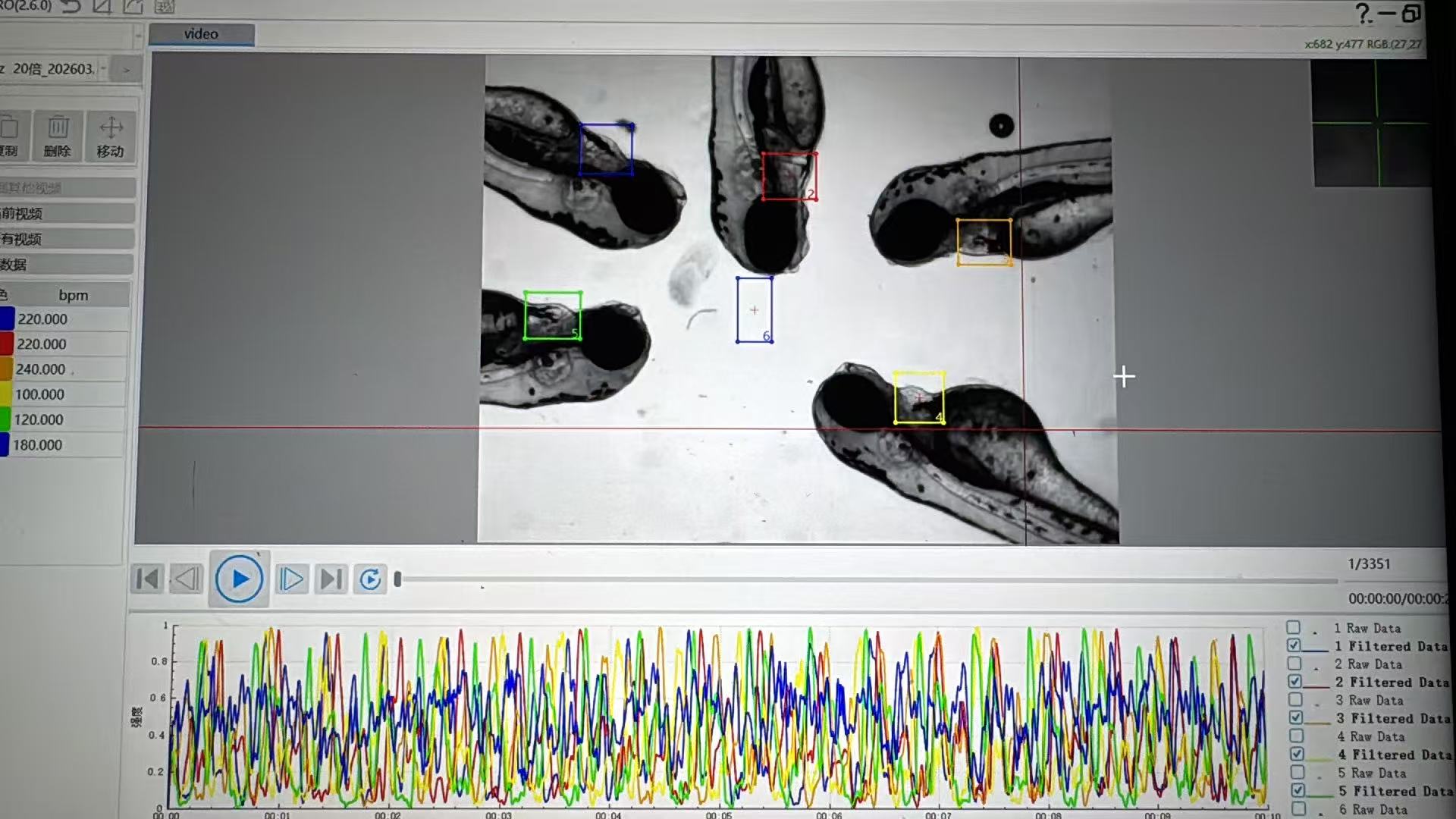
For a cardiovascular program, early screening is not simply a matter of recording whether a zebrafish embryo looks normal after exposure. The useful question is whether a compound changes circulation in a way that deserves closer study. A Zebrafish Blood Flow Analysis System gives a screening service a repeatable way to capture that question: acquire comparable images, define endpoints before scoring, and turn observations into a traceable review package.
From a drug discovery perspective, zebrafish cardiovascular assays are valuable because they let teams examine living circulation while keeping the workflow practical for early decisions. A well-planned Zebrafish Blood Flow Analysis System does not replace mammalian pharmacology, clinical assessment, or a full safety program. It can, however, help a team decide which findings merit escalation and which assay conditions need another look.
What the system adds to a cardiovascular screening service
Blood-flow analysis connects the imaging step to an operational service workflow. Rather than storing a collection of videos with loosely written notes, the service can define a plate map, exposure schedule, imaging window, control set, and scoring rules before the first run. The Zebrafish Blood Flow Analysis System then supports a consistent record of circulation-related observations across test articles and repeat runs.
That record is especially useful when a compound creates a subtle phenotype. Heart rhythm, chamber movement, vessel appearance, red-cell movement, edema, body orientation, and embryo viability may all matter, but they do not carry the same meaning in every protocol. Cardiovascular endpoints should be selected around the research question, not copied from a generic checklist. If the aim is early drug screening, a service should define which endpoint triggers repeat imaging, concentration review, or a follow-on assay.
The practical value comes from comparing like with like. The imaging setup, developmental stage, temperature handling, anesthetic exposure, orientation method, and illumination plan should be documented for each run. Small changes in those conditions can influence an apparent blood-flow difference. A Zebrafish Blood Flow Analysis System is most helpful when it sits inside that controlled workflow rather than being treated as a camera alone.
A representative service workflow
1. Translate the sponsor question into observable endpoints
A sponsor may ask whether a series has a cardiovascular liability signal, whether two formulations behave differently, or whether a lead can move into a more resource-intensive study. The service team should convert that broad request into a protocol question: which animals, concentrations, timepoints, controls, and circulation measures can inform the next decision? This is where a Zebrafish Blood Flow Analysis System brings structure to drug screening services.
For example, a screening plan can predefine a comparison of vehicle controls, reference conditions when appropriate, and blinded image review. The plan should also state whether the output is descriptive, semi-quantitative, or quantitative. Clear language matters: a screening result may identify a signal for further investigation; it does not by itself establish a mechanism or a human outcome.
2. Standardize acquisition before trying to increase throughput
Our first instinct is often to push throughput as high as possible, but inconsistent larval orientation can quietly undermine circulation interpretation. A reliable service balances plate capacity with image quality, focus stability, and traceability. Automated orientation or a defined mounting procedure can help, provided the team verifies that the method suits its own strain, developmental window, and endpoint.
Use a run sheet that records the operator, plate identity, imaging settings, exposure timing, and any deviations. When a reviewer sees an unusual circulation pattern, that context is as important as the image itself. This approach makes a Zebrafish Blood Flow Analysis System more useful across projects because reviewers can distinguish a possible compound effect from an acquisition issue.
3. Pair blood-flow observations with contextual morphology
Blood flow should not be interpreted in isolation. A decrease in visible movement may occur alongside developmental delay, edema, poor orientation, general toxicity, or an image-quality limitation. Conversely, a morphology-first review can miss a functional change that becomes clearer in the circulation record. The service workflow should therefore connect cardiovascular observations with viability and morphology notes without overstating causality.
We started out treating a single readout as the most direct indicator, but a combined review of circulation, heart appearance, and general condition often gives the more useful triage picture. If conditions differ between imaging sessions, the calculation needs adjustment or the run may require repeat acquisition. That measured approach helps drug screening teams communicate what the data do and do not support.
How to make the output useful for sponsors
For contract research and in-house discovery teams, the deliverable should be easy to interrogate. A good report can include the study objective, test-item handling notes, plate and sample identifiers, predefined endpoints, representative images or clips, reviewer annotations, deviation notes, and a decision-oriented summary. The Zebrafish Blood Flow Analysis System supplies the imaging and analysis backbone; the service design supplies the chain of custody and interpretation boundaries.
That distinction is important for cardiovascular work. A conclusion such as “a signal was observed under the tested conditions” is more credible than a broad claim about safety or efficacy. When a result is equivocal, a well-run service should say so, explain the limiting condition, and propose the next appropriate experiment. Mild uncertainty is not a weakness in an early screen; it is often the information that guides better study design.
Questions to ask when evaluating a service workflow
- Can the provider help define cardiovascular endpoints that match the sponsor’s decision point?
- How are zebrafish developmental stage, handling, orientation, and imaging settings documented?
- What controls and repeat criteria are set before drug screening begins?
- How does the workflow separate image-quality concerns from biological observations?
- Can the final record support internal review and a later follow-on study?
These questions shift the discussion beyond a feature list and toward reproducibility. Hunter Lab focuses on zebrafish instruments and applications, and its cardiovascular-analysis experience can inform pre-purchase protocol discussions and training. The exact scope should be confirmed against the project’s assay, site requirements, and intended use.
Where the approach fits in a broader development plan
A Zebrafish Blood Flow Analysis System is most effective as one part of a staged evidence plan. It can support early prioritization, method development, and comparative drug screening when the protocol is transparent and the endpoints are relevant. Findings may then be paired with chemistry, in vitro work, pharmacokinetics, or other models according to the program’s needs.
For cardiovascular drug screening services, the central benefit is not a promise of a single answer. It is a disciplined way to see circulation-related changes early, preserve the context behind each observation, and make the next experimental choice with more confidence. Start with the decision you need the screen to support, then configure the imaging and review process around that decision.








