
 EN
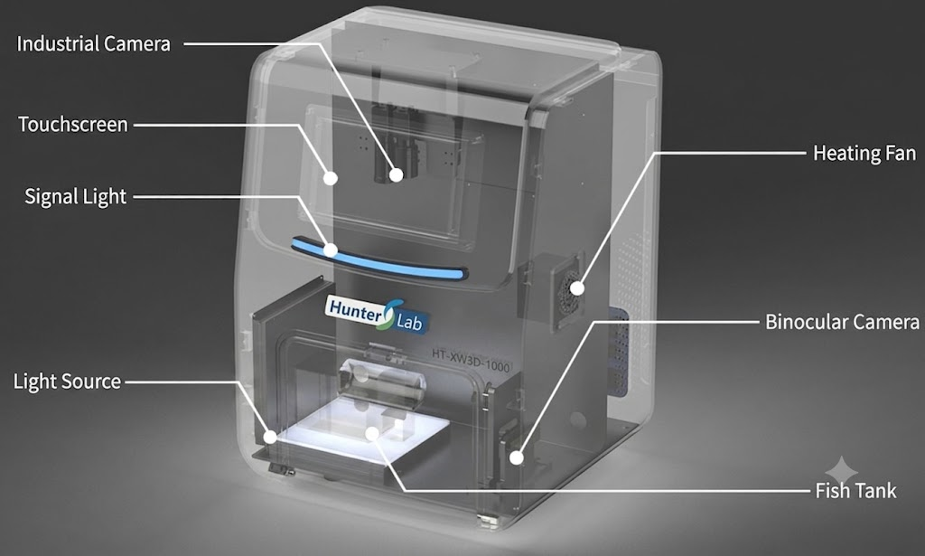
Advanced zebrafish fluorescence imaging systems feature automated multi-well scanning.
A Zebrafish Fluorescence Imaging System becomes valuable when it turns a crowded plate and a busy imaging schedule into a documented, repeatable workflow. For a core facility manager, the appeal of automated multi-well scanning is not simply that a stage moves faster. The practical goal is to acquire comparable fluorescence images across wells while preserving the experimental details that explain what each image means.
Zebrafish embryos are optically accessible, which is one reason live fluorescence imaging can reveal changing cells, tissues, and organ-level signals during development. Yet that accessibility creates its own operational challenge: a screen may involve many embryos, multiple conditions, several fluorescence channels, and time-sensitive handling. A Zebrafish Fluorescence Imaging System with automated scanning can make that complexity manageable only when the acquisition plan, plate layout, focus rules, and quality controls are set before the first well is captured.
Why multi-well scanning changes the daily workflow
In a manual workflow, an operator chooses a well, centers an embryo, adjusts focus, captures an image, and repeats. This approach can be useful for exploratory work, but it becomes hard to scale when the same study must be repeated across a plate or across several days. Automated multi-well scanning coordinates well positions with a defined capture sequence. Depending on the configuration, that sequence may include brightfield context, fluorescence channels, autofocus or focus offsets, and standardized file naming.
The efficiency benefit is therefore broader than fewer clicks. It can reduce avoidable variation in how fields are selected, clarify which settings were used, and leave the operator free to review exceptions rather than perform every navigation step. That does not mean automated acquisition is inherently more accurate. A poorly focused or poorly normalized automated scan can reproduce the same error very efficiently. The longer answer depends on whether the lab validates the workflow against representative specimens and controls.
What to evaluate in a Zebrafish Fluorescence Imaging System
Before comparing platforms, define the decision in terms of the assay rather than a generic request for high-throughput imaging. The following questions help establish whether automated multi-well scanning is a good fit.
- Plate and sample format: Confirm the well geometry, imaging area, embryo orientation expectations, and whether the assay needs one field or multiple positions per well.
- Signal and channel plan: Specify fluorophores, expected signal range, exposure constraints, and whether brightfield images are needed to interpret the fluorescence result.
- Focus strategy: Test autofocus or fixed focus offsets on the specimens that matter. Focus behavior often changes with well depth, mounting medium, developmental stage, or orientation.
- Metadata discipline: Decide how plate map, treatment, developmental timing, channel, exposure, and operator notes will travel with the images.
- Review path: Plan how failed focus, debris, motion, or low-signal wells will be flagged for human review instead of silently entering analysis.
A Zebrafish Fluorescence Imaging System should support this decision framework without forcing the research team to claim more standardization than it has actually achieved. In multi-user facilities, consistency of the SOP can matter as much as a feature list. A new user should know which settings are fixed, which are assay-specific, and when to stop an acquisition for review.
Designing an automated scan that produces usable data
Start with a plate map that serves analysis
Multi-well scanning is most useful when the plate map already anticipates analysis. Include untreated controls, positive controls where they are appropriate, replicate logic, and any wells reserved for focus or exposure checks. Avoid treating a plate as a neutral container. Edge effects, timing differences, and handling variation can all influence a fluorescence readout, so the acquisition record should make those sources visible.
Normalize before scaling
For each channel, establish a condition in which the signal is detectable without routinely clipping the brightest regions. Then test the settings across a small but representative set of wells. Researchers sometimes assume that higher resolution or more frames automatically improve a result. Actually, an image only helps if its quality is consistent enough for the intended measurement. A high-throughput workflow should therefore document exposure, gain where applicable, focus method, and any post-acquisition processing rules before a full plate is run.
Use automation to protect reproducibility
The most useful automated workflows make routine steps explicit. A scan order can be fixed; filenames can encode plate and well identity; image folders can be organized by assay run; and exception wells can be reviewed against the same criteria. Those details matter when an experiment is repeated weeks later or transferred between operators. In practice, a Zebrafish Fluorescence Imaging System earns its place when it reduces undocumented choices, not when it merely produces a larger image archive.
Balancing high-throughput goals with image quality
High-throughput screening creates a real trade-off. Shorter acquisition times can increase throughput, while multiple focal planes, repeated channels, and manual review can increase confidence in the result. Neither side wins by default. For a pilot study, the lab may accept a slower scan to establish a reliable phenotype definition. For a mature assay, the same lab may prioritize standardized automation and sample volume once the decision thresholds are validated.
Most teams benefit from keeping a small verification set in every run. It can include known controls, a subset reviewed manually, and checks for background, focus, and signal saturation. If the verification set begins to diverge from the automated analysis, investigate the protocol, sample preparation, and imaging settings before concluding that the biology changed. This is particularly important when results will be compared across days or operators.
A commissioning checklist for automated multi-well scanning
- Define the biological question and the fluorescence feature to be measured.
- Test plate compatibility, well positioning, and specimen orientation with representative samples.
- Set channel, exposure, and focus rules using controls rather than a single visually attractive field.
- Run a pilot plate and review image quality well by well before scaling the protocol.
- Document acceptance criteria, exception handling, and metadata requirements in an SOP.
- Repeat the pilot with another operator to identify training gaps and inter-operator variation.
When a lab is considering a Zebrafish Fluorescence Imaging System, pre-purchase application consultation can be useful for testing these workflow assumptions against the proposed assay. The key is to ask for an evidence-based discussion of the sample format, acquisition logic, and validation plan, rather than relying on generic throughput promises.
Making the selection decision
The right platform depends on whether the bottleneck is navigation, focus consistency, data organization, image analysis, or operator capacity. A small discovery project may need flexible manual control alongside light automation. A screening program with a validated plate-based assay may benefit more from repeatable scan templates and clear quality-control gates. In both cases, the practical measure of efficiency is whether the team can make defensible comparisons from the images it collects.
Automated multi-well scanning is a meaningful advance because it can make imaging routines more repeatable and reviewable. Used carefully, a Zebrafish Fluorescence Imaging System helps teams move from isolated images toward a controlled acquisition process without losing sight of the biological and technical checks that give fluorescence data its value.








